There is an old clichØ that Access is Success . Unlike other aspects of dentistry, root canal treatment is carried out with little visual guidance; therefore, the difficulties that are likely to be encountered need to be considered. An assessment of the following features can be made after visual examination of the tooth, and study of a pre-operative periapical radiograph taken with a paralleling technique:
The external morphology of the tooth. The architecture of the tooth s root canal
system. The number of canals present. The length, direction and degree of curvature
of each canal. Any branching or division of the main canals. The relationship of the canal orifice(s) to the pulp chamber and to the external surface of the tooth.
The presence and location of any lateral canals.
The position and size of the pulp chamber and its distance from the occlusal surface.
Any related pathology.
Before commencement of root canal treatment, the tooth must be prepared as follows:
All caries and any defective restorations should be removed and made good. The tooth should be protected against fracture during treatment.
The tooth should be capable of isolation. The periodontal status should be sound, orcapable of resolution. It may be prudent to commence access cavity
preparation before isolating the tooth with rubber dam in order that the anatomical landmarks, tooth inclination and other helpful features are not lost. It is, of course, crucial that the root canal does not become contaminated during either access preparation or canal instrumentation, and the tooth should be isolated in an aseptic field as soon as possible.
If there is a danger of fracture of the coronal tooth structure, the cuspal height should be reduced to prevent this. If the loss of coronal tissue is extensive, there may be a need to provisionally restore the tooth with a temporary crown, copper ring or an orthodontic band. It is, however, not always necessary to restore the tooth before carrying out endodontic procedures. Provided the tooth will anchor a rubber dam, the canals can be isolated from the oral cavity and a temporary seal can be placed over the canals, this will be sufficient.
The objectives of access cavity preparation are to:
Remove the entire roof of the pulp chamber so that the pulp chamber can be debrided.
Enable the root canals to be located and instrumented by providing direct straight line access to the apical third of the root canals, as illustrated in Figure 6.17. Note that the initial access cavity may have to be modified during treatment to achieve this.
Enable a temporary seal to be placed securely in order to withstand any displacing forces.
Conserve as much sound tooth tissue as possible and as is consistent with treatment objectives.
The subsequent restoration of the tooth should always be considered first. If the tooth is not heavily restored then only the amount of coronal tissue sufficient for the successful completion of the root canal treatment should beremoved. However, if the tooth is already compromised and will require some form of cuspal coverage restoration, an onlay or a crown, then it may be practical to reduce the cusp height, particularly mesiobuccally in molars, to enable better visualisation of the pulp chamber. If access to the back of the mouth is difficult, it is again reasonable to consider reducing the marginal ridge of the tooth concerned to achieve this (Fig. 18), or perhaps gain access through the mesiobuccal wall. Unless the root treatment is successful, any further restoration to the tooth will be put at risk.
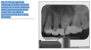
Before beginning the access cavity preparation, it is wise to check the depth of the preparation by aligning the bur and handpiece against the radiograph, in order to note the position and depth of the roof of the pulp chamber in relation to the length of the bur in the handpiece (Fig. 19). Particular note should be made of the position of the largest pulp horn.
The stages of access cavity preparation may be summarised as follows:
1. The initial entry is made with a tungsten carbide or diamond bur in a turbine handpiece and the outline form completed as required. The bur is advanced towards the pulp horns until the roof of the pulp chamber is just penetrated. (Note particularly that in a molar tooth the bur approaches the tooth from the mesial and from the buccal. Thus the access cavity is cut in the mesiobuccal segment of the occlusal surface.)
2. At this point, the rubber dam should be applied if it is not already in place.
3. The removal of the entire roof of the pulp chamber, and the tapering of the walls, is now carried out with a safe-tipped endodontic access bur, as described in Part 5. (Stages 1 and 3 are illustrated in the diagrams in Fig. 20.)
4. The walls of the pulp chamber may now be gently flared out towards the occlusal surface. The end result should be a gentle funnelshape, with the larger diameter at the occlusal surface. The safe tip of the bur will be felt passively following the contours of the floor of the pulp chamber.
5. Any remaining pulp tissue and debris is cleared with an excavator from the floor of the pulp chamber and the canal orifices.
6. The access cavity should be flushed with a solution of sodium hypochlorite to remove any residual debris.
7. The canal orifices may be located with a DG 16 endodontic probe. Any alteration to the access cavity outline form may now be undertaken to ensure a direct line of approach to the canal orifices. Any sclerotic or secondary dentine surrounding the canal orifices may be removed with a CT4 tip in a piezo-electronic ultrasonic machine.
8. Once the canal orifices have been identified, the preparation of the coronal part of the root canals should be commenced. Depending upon the operator s preferred technique, either Gates Glidden burs or nickel-titanium orifice shapers, should be employed. Copious irrigation is necessary, together with the use of a canal lubricant containing EDTA. These techniques are described in Part 7.
