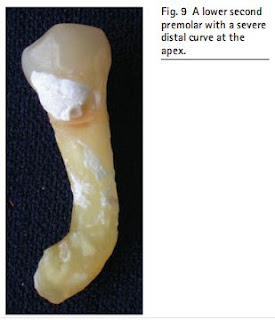
This tooth is similar to the first premolar, except that the incidence of a second canal is very much lower. One study stated this to be 12%. Another study revealed that only 2.5% had two apical foramina. Consequently, it is a much easier tooth to treat compared with the mandibular first premolar, unless the radiograph reveals a sharp distal curve at the apex as shown in the extracted tooth at Figure 9.
